skip to Main Content

Sitagliptin Treatment at the Time of Hospitalization Was Associated With Reduced Mortality in Patients With Type 2 Diabetes and COVID-19: A Multicenter, Case-Control, Retrospective, Observational Study

Sebastiano Bruno Solerte, Francesca D’Addio, Roberto Trevisan, Elisabetta Lovati, Antonio Rossi, Ida Pastore, Marco Dell’Acqua, Elio Ippolito, Cristiana Scaranna, Rosalia Bellante, Silvia Galliani, Alessandro Roberto Dodesini, Giuseppe Lepore, Francesca Geni, Roberta Maria Fiorina, Emanuele Catena, Angelo Corsico, Riccardo Colombo, Marco Mirani, Carlo De Riva, Salvatore Endrio Oleandri, Reza Abdi, Joseph V. Bonventre, Stefano Rusconi, Franco Folli, Antonio Di Sabatino, Gianvincenzo Zuccotti, Massimo Galli1 and Paolo Fiorina

September, 2020

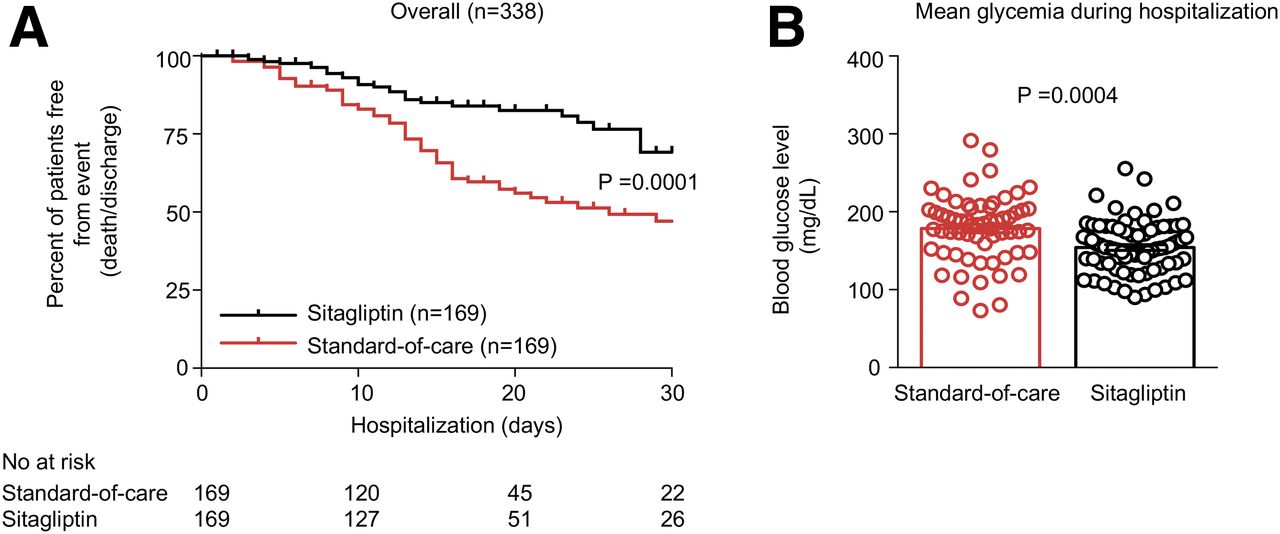
Poor outcomes have been reported in patients with type 2 diabetes and coronavirus disease 2019 (COVID-19); thus, it is mandatory to explore novel therapeutic approaches for this population.


Lymph node fibroblastic reticular cells deposit fibrosis-associated collagen following organ transplantation

Xiaofei Li et. al.

August, 2020

Although the immune response within draining lymph nodes (DLNs) has been studied for decades, how their stromal compartment contributes to this process remains to be fully explored. Here, we show that donor mast cells were prominent activators of collagen I deposition by fibroblastic reticular cells (FRCs) in DLNs shortly following transplantation.


Regulatory CD8 T cells that recognize Qa-1 expressed by CD4 T-helper cells inhibit rejection of heart allografts

John Y. ChoiSiawosh K. EskandariSongjie CaiIna SulkajJean Pierre AssakerHazim AllosJuliano AlHaddadSaif A. MuhsinEman AlhussainAmr MansouriMelissa Y. YeungMarc A. J. SeelenHye-Jung Kim, Harvey Cantor, and Jamil R. Azzi

March, 2020

Induction of longstanding immunologic tolerance is essential for survival of transplanted organs and tissues. Despite recent advances in immunosuppression protocols, allograft damage inflicted by antibody specific for donor organs continues to represent a major obstacle to graft survival.


Follicular regulatory T cells control humoral and allergic immunity by restraining early B cell responses

Rachel L. Clement, Joe Daccache, Mostafa T. Mohammed, Alos Diallo, Bruce R. Blazar,
Vijay K. Kuchroo, Scott B. Lovitch, Arlene H. Sharpe and Peter T. Sage.

October, 2019

Follicular regulatory T (TFR) cells have specialized roles in modulating follicular helper T (TFH) cell activation of B cells. However,
the precise role of TFR cells in controlling antibody responses to foreign antigens and autoantigens in vivo is still unclear due to a
lack of specific tools.


FoxP3 and Ezh2 regulate Tfr cell suppressive function and transcriptional program

Shenda Hou, Rachel L. Clement, Alos Diallo, Bruce R. Blazar, Alexander Y. Rudensky, Arlene H. Sharpe, and Peter T. Sage.

January, 2019

Follicular regulatory T (Tfr) cells are a regulatory T cell subset that controls antibody production by inhibiting T follicular
helper (Tfh)–mediated help to B cells. Tfh and Tfr cells possess opposing functions suggesting unique programming. Here we
elucidated the transcriptional program controlling Tfr suppressive function.


Back To Top